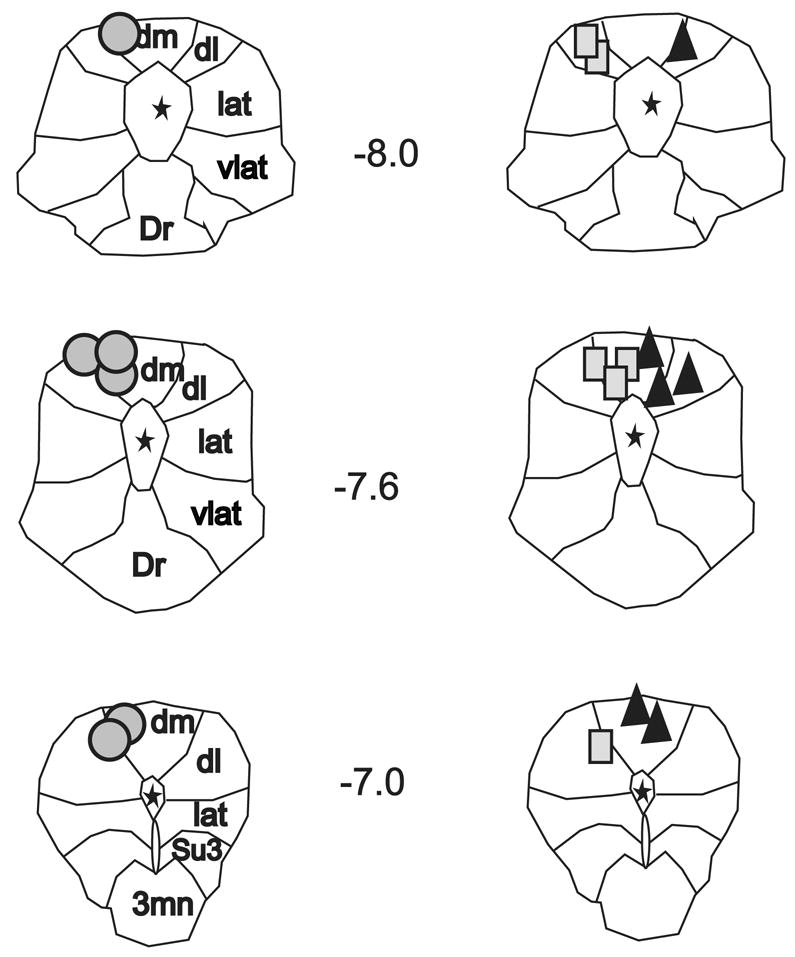
Fig. 3. Reconstructed dorsal PAG stimulation sites.

Outline of the rostral, middle, and caudal columns of the PAG from Paxinos & Watson’s Rat Brain Stereotaxic Atlas (35). Reconstructed stimulation sites from all animals are organized by experimental group; circles indicate PAG stimulation sites recovered from muscimol microinjection studies; triangles indicate sites from kynurenic acid studies; and rectangles indicate sites from aCSF studies. Note, for illustration purposes all stimulation sites from kynurenic acid experiments are shown on the right side. Numbers refer to midbrain location relative to bregma. Asterisks indicated location of central aqueduct. Dm, dorsomedial column; dl, dorsolateral column; lat, lateral column; vlat, ventrolateral column; Dr, dorsal raphe; Su3, supraoculomotor nucleus; 3mn, oculomotor nucleus.
