
FIGURE 3.
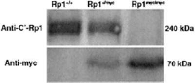
Western-blot analysis of wild-type Rp1 and mutant Rp1-myc protein expression. Total retinal proteins were extracted from 4-week-old Rp1+/+Rp1+/myc, and Rp1myc/mycmice. Equal amounts of total protein from each genotype were electrophoresed and transferred to polyvinylidene difluoride (PVDF) membrane and then probed with either antibodies to the C terminus of Rp1 (top: anti-C′-Rp1 antibody) or anti-myc (bottom). The normal mouse Rp1 protein (˜240 kDa) was detected in Rp1+/+and Rp1+/mycmice, but not in Rp1myc/mycmice. An ˜70-kDa protein corresponding to the predicted size of the mutant Rp1-myc protein was present in Rp1+/mycand Rp1myc/mycmice, but absent in Rp1+/+mice.