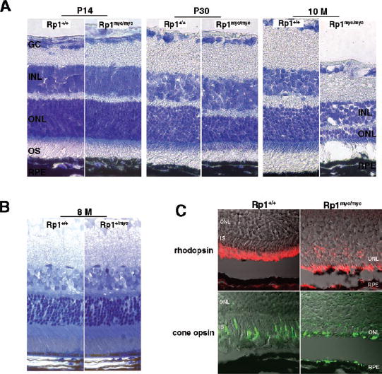
FIGURE 8.

Progressive degeneration of photoreceptors in the Rp1myc/mycmice. (A)The extent of photoreceptor loss in Rp1myc/mycmice was estimated by observing the thickness of the outer nuclear layer (ONL) in sections of frozen retina from mutant and control mice at different ages. Lens-optic nerve sections were stained with Richard-son stain. At age P14, the thickness of the ONL was comparable between Rp1myc/mycand wild-type littermates (left). By 30 days of age, retinas in Rp1myc/mycmice showed significant degenerative changes. The thickness of the ONL was reduced by approximately two to three rows compared with wild-type littermate control animals (middle). By 10-months of age, only two rows of nuclei remained in the retinas of Rp1myc/mycmice (right). (B)Toluidine blue staining of Epon-embedded retinal sections from 8-month-old Rp1+/+and Rp1+/mycmice shows that the thickness of ONL and the length of outer segments did not differ significantly between Rp1+/+and Rp1+/mycmice. (C) Immunofluorescence and Nomar-ski confocal images of retinas in 10-month-old Rp1+/+and Rp1myc/mycmice. Antibodies against rhodopsin (red, top)and red/greencone opsin (green, bottom)show that both rods and cones remain in the degenerated Rp1myc/mycretinas. Mislocalization of both proteins to the inner segment and ONL was detected in the mutant mice.
