
Figure 1. Neonatal monocytes isolated from fresh cord blood are deficient at MHC-II antigen processing and presentation.
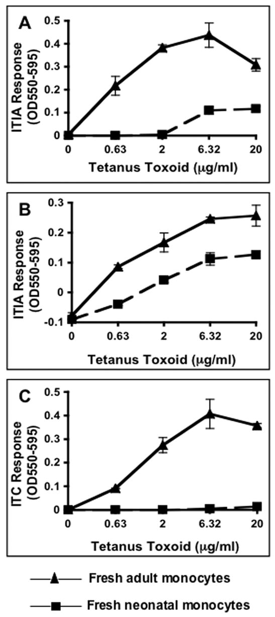
Negatively selected monocytes isolated from fresh HLA-matched cord and adult blood was incubated for 24 h with antigen and appropriate HLA-restricted T hybridoma cells. Supernatants were assessed for IL-2 content by a CTLL-2 proliferation assay that was monitored with Alamar blue, an indicator dye. A & B) HLA-DR1 (allele DRB1*0101) neonatal and adult monocytes incubated with tetanus toxoid and ITIA T hybridoma cells. C) HLA-DR4 (allele DRB1*0401) neonatal and adult monocytes incubated with tetanus toxoid and ITC T hybridoma cells. Data points are means of triplicate samples +/− S.D.