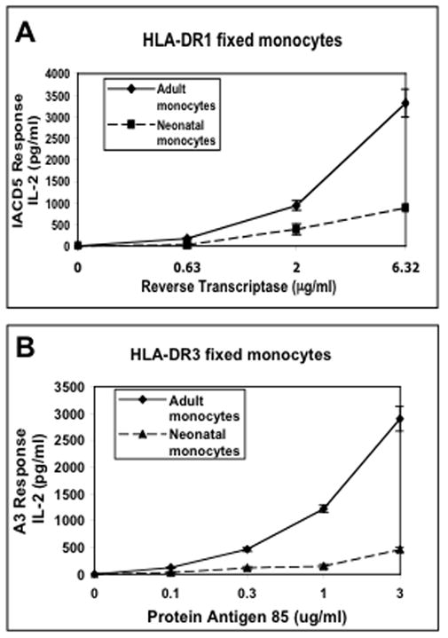
Figure 4. Fixed neonatal monocytes are also defective in MHC-II antigen presentation.

Negatively-selected monocytes isolated from HLA-DR1 or HLA-DR3 cord or adult blood-derived PBMCs were incubated for 24 h with reverse transcriptase and antigen 85 respectively. Cells were washed, fixed in 1% paraformaldehyde (to prevent secretion of any soluble factors) and then incubated for an additional 24 h with A) IACD5 T hybridoma cells or B) A3 T hybridoma cells. Supernatants were assessed for IL-2 content by IL-2 ELISA. Data points are means of triplicate samples +/− S.D.
