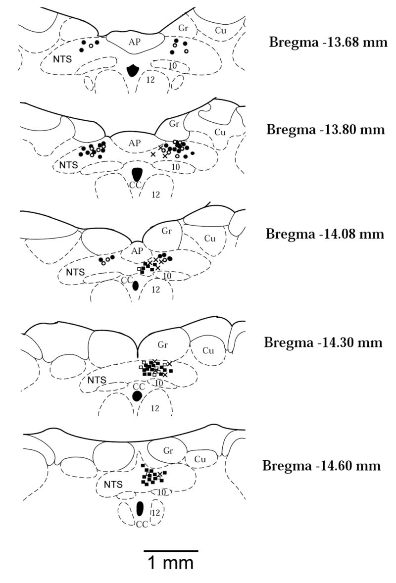
Fig. 1.

Anatomic locations of recorded neurons plotted on standard coronal sections. Barosensitive neurons in which discharge was decreased (●, 38 sites) or did not respond (○, 16 sites) to epicardial capsaicin; Chemosensitive neurons in which discharge was increased (▪, 33 sites) or did not respond (□, 9 sites) to epicardial capsaicin; Neurons in which discharge was increased by both baroreceptor and chemoreceptor stimulation (×, 12 sites). AP, area postrema; CC, central canal; Cu, cuneate nucleus; Gr, gracile nucleus; NTS, nucleus tractus solitarius; 10, motor nucleus of vagus nerve; 12, nucleus of hypoglossal nerve.
