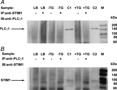
Figure 9. Immunoprecipitation of STIM1 and PLC-γ1.
(A) Cell lysates (100 μg) or lysis buffer only (LB) were immunoprecipitated (IP) with anti-STIM1 antibody (+) [or PBS as a negative control (−)] and the immunoprecipitates were analysed by immunoblotting (IB) with anti-PLCγ1 antibody. H4IIE cells were either untreated (−TG) or treated (+TG) with 1 μM thapsigargin and cell lysates were prepared using Ca2+-supplemented (5 mM free Ca2+) lysis buffer. Positive controls included non-immunoprecipitated lysates (10 μg) from cells untreated (C1) or treated (C2) with 1 μM thapsigargin. No STIM1-immunoprecipitated 150 kDa PLC-γ1 band was observed in either the untreated or thapsigargin treated cell lysates. Molecular mass markers (M) are indicated to the right-hand side of the Figure. The results are representative of two independent experiments. (B) Cell lysates (100 μg) or lysis buffer only (LB) were immunoprecipitated (IP) with anti-PLC-γ1 antibody (+) [or PBS (−)] and the immunoprecipitates were analysed by immunoblotting (IB) with anti-STIM1 antibody. No PLCγ1-immunoprecipitated 85 kDa STIM1 band was observed in either the untreated or thapsigargin-treated cell lysates. The results are representative of two independent experiments. The band at approx. 75 kDa present in the IP(+) samples, including the lysis buffer only sample, is most probably non-denatured anti-PLCγ1 immunoprecipitating antibody. The faint band at approx. 95 kDa present in both IP(−) and IP(+) samples, including the lysis buffer only samples, is most probably an artefact due to the Protein A/G Plus agarose beads.

