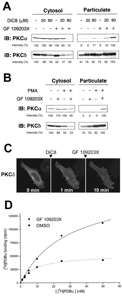
Figure 6. GF 109203X enhances DiC8- and PMA-induced translocation of endogenous PKC and [3H]PDBu binding to the cytosolic fraction prepared from PKCα–GFP-overexpressing cells.
(A) HL-60 cells were pretreated with 0.5 μM GF 109203X for 20 min and then stimulated with 20 or 80 μM DiC8 for 5 min. (B) HL-60 cells were pretreated with 1 μM GF109203X for 20 min and then stimulated with 10 nM PMA for 15 min. Subcellular fractions were obtained as described in the Experimental section and subjected to SDS/PAGE, followed by immunoblotting (IB) with anti-PKCα and anti-PKCδ antibodies. Densitometric analysis was performed using Scion Image software. In the cytosolic fraction the intensity of the protein bands is shown as a percentage of that of the control cells. In the particulate fraction the intensity of the protein bands is shown as a percentage of that of the 80 μM DiC8-plus-GF 109203X (A)- or PMA-plus-GF 109203X (B)-treated cells. Results are representative of at least three separate experiments. (C) HeLa cells transiently expressing PKCδ–GFP were pretreated with 10 μM DiC8 for 1 min, and then treated with 1 μM GF 109203X for 9 min. Results are representative of at least three separate experiments. (D) The PDBu binding was evaluated by the method described in the Experimental section in the presence of DMSO or 10 μM GF 109203X. The expression of PKCα–GFP was confirmed by immunoblotting with anti-GFP and anti-PKCα antibodies. Results are representative of at least three separate experiments, with duplicate determinations in each experiment.

