
Fig. 6.
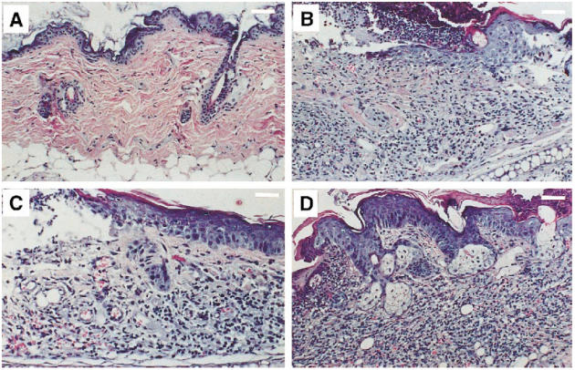
Dermatohistopathology in TCRα+ MRL and TCRα− MRL mice. (A) Some young TCRα− MRL/lpr animals had normal skin pathology, as seen in this neck specimen from a 24-week-old animal (scale bar, 200 μm). (B) In contrast, 24-week-old TCRα+ MRL/lpr animals consistently developed ulceration, hyperkeratosis, acanthosis, and mononuclear cell infiltration into the dermis and epidermis by 24 weeks old (here, a neck specimen; scale bar, 200 μm). (C) Many 24-week-old TCRα− MRL mice, however, developed subclinical ulceration, fibrosis, and mild dermal infiltration (here, a TCRα− MRL/lpr neck; 50–70% of TCRα− MRL/lpr versus 20–30% of TCRα− MRL +/+ animals developed such lesions; scale bar, 125 μm), which soon developed into (D) florid lesions, such as in this 28-week-old TCRα− MRL +/+ neck, which contained striking infiltrates, as well as hyperkeratosis, acanthosis, and dermal fibrosis (scale bar, 200 μm).