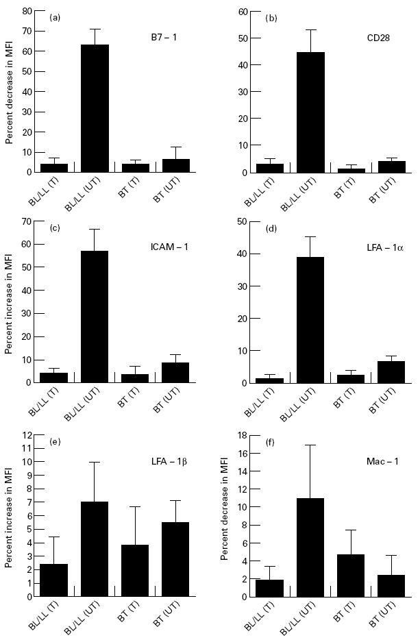
Fig. 3.

Expression of B7-1, CD28, intercellular adhesion molecule-1 (ICAM-1), LFA-1α, LFA-1β and Mac-1 on freshly isolated lymphocytes of leprosy patients. The lymphocytes were stained using MoAbs against the costimulatory molecules (5 ng/ml) and secondary FITC-labelled antibodies. Data shown are represented as percent change in the mean fluorescence intensity (MFI) of the costimulatory molecules expressed on the surface of the lymphocytes obtained from the leprosy patients (mean ± s.d.; borderline leprosy (BL)/lepromatous leprosy (LL) treated (T), n = 3; BL/LL untreated (UT), n = 6; borderline tuberculoid (BT) leprosy treated (T), n = 7; BT untreated (UT), n = 3), compared with the expression of costimulatory molecules on the lymphocytes of healthy subjects (mean ± s.d.; n = 7). No staining was viewed in the controls containing isotype-matched MoAbs.
