
Fig. 4.
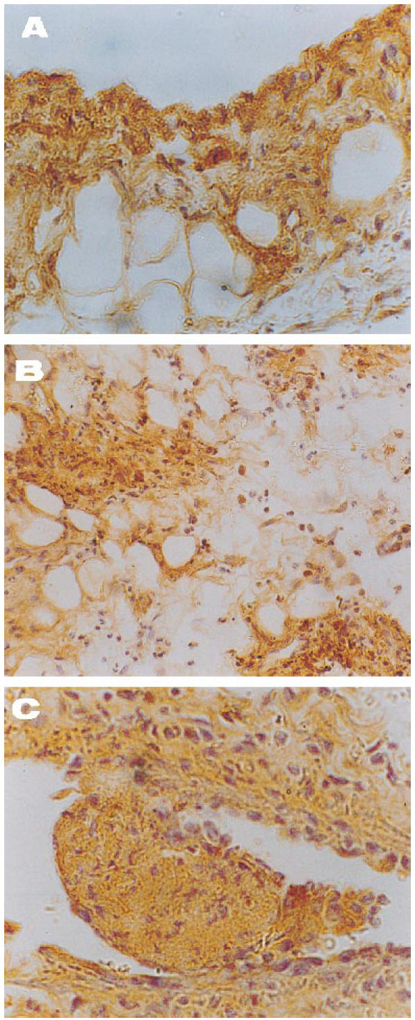
Pattern of localization of IL-8 and MCP-1 in synovial tissues of rabbits with acute antigen arthritis. Both anti-chemokine antibodies displayed a similar distribution in the injured tissue, preferentially staining the lining layer, areas of neutrophil infiltration and endothelium. The figure shows representative findings of untreated specimens employing immunoperoxidase techniques. (A) Binding of anti-IL-8 antibody to synovial lining (×400). (B) The strong IL-8 staining of the interstitium is limited to granulocyte accumules (×200). (C) Organized thromboses surrounded by vasculitic damage are typical of this experimental disease. Note the striking positivity for MCP-1 within the thrombosis and around the vessel wall (×400).