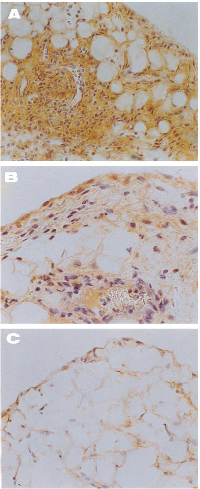
Fig. 5.

Immunolocalization of IL-8 in joint tissue from arthritic rabbits. (A) Intense and diffuse positive signal in an untreated rabbit, including a mural thrombosis and perivascular infiltration (×200). (B) Note the mild staining of superficial synovial cells and infiltrates in a tenidap-treated animal (×400). (C) A demonstrably healthy synovium where IL-8 was almost absent (×200). There was no staining in the negative controls included in each experiment (not shown).
