
Fig. 6.
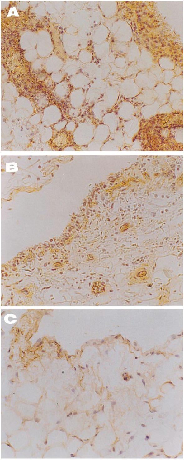
Immunolocalization of MCP-1 in joint tissue from arthritic rabbits. (A) An untreated specimen shows lining hyperplasia markedly positive for MCP-1 with several infiltrates which also bound the specific antibody (×200). (B) In a tenidap-treated synovium a diminution of the former signal is evident, with slight staining of endothelium and lining cells (×200). (C) Healthy control tissue, where minimal binding of anti-MCP-1 is seen at the lining layer (×200). There was no staining in the negative controls included in each experiment (not shown).