
Fig. 2.
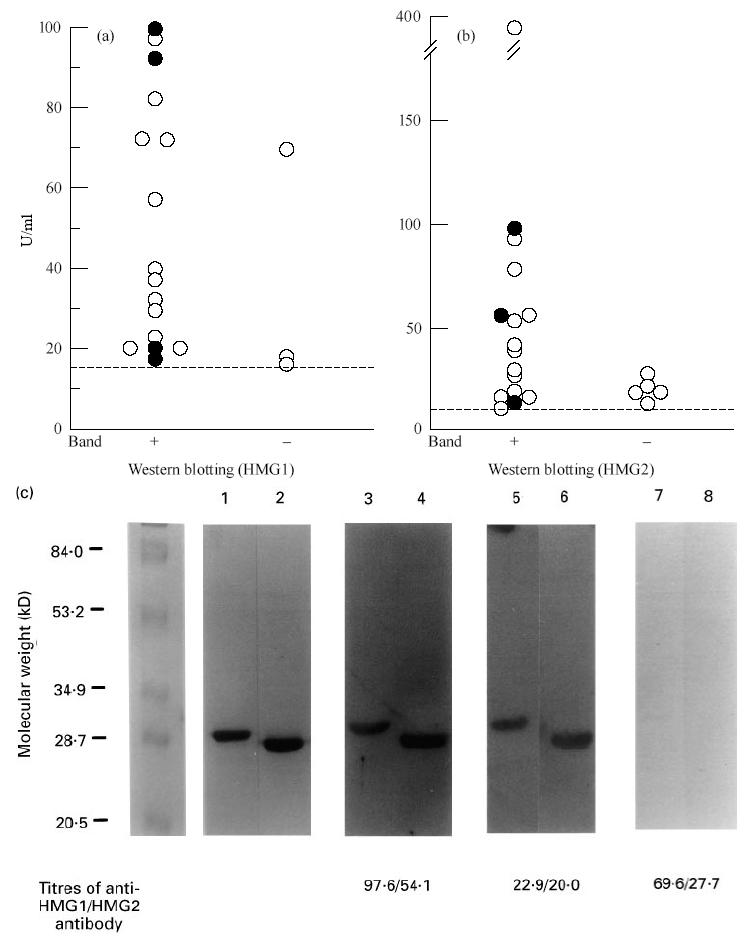
Detection of antibodies to HMG1/HMG2 by ELISA and Western blotting. (a) The 19 sera positive for anti-HMG1 antibody by ELISA were tested by Western blotting using HMG1 as antigen. (b) The 20 sera positive for anti-HMG2 antibody by ELISA were tested by Western blotting using HMG2 as antigen. Closed and open circles represent individual patients with refractory and non-refractory types of UC, respectively. (c) Western blotting analysis of anti-HMG1/HMG2 antibody. Antigenic substrates were 1 μg porcine HMG1 (lanes 1, 3, 5 and 7) or HMG2 (lanes 2, 4, 6 and 8). Lanes 1 and 2, SDS–PAGE stained by Fast stain (Zoion, Allestone, MA); lanes 3–8, sera of UC patients. The titres of anti-HMG1/HMG2 antibody of these sera are indicated below the lanes.