
Fig. 9.
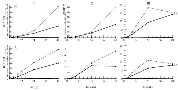
Secretion of IL-8 by three donor retinal pigment epithelial (RPE) cell cultures (I, II, III) after stimulation with IL-1β from the apical side (a) or the basal side (b). RPE cell lines were seeded on transwell filters at a concentration of 1.6 × 10⁵ cells/cm2 and cultured for at least 19 days in Iscove's modified Dulbecco's medium (IMDM) supplemented with 1% fetal calf serum (FCS) and were used when net trans-epithelial resistance (TER) was at least 20 Ω·cm2. Before stimulation monolayers were cultured for 24 h in serum-free medium. Filters were stimulated from either the upper or lower compartment with IL-1β at a final concentration of 100 U/ml. Data are expressed as absolute amounts of IL-8 per filter and are expressed as the mean of two filters. Statistical analysis of the log transformed data revealed a significant difference between the upper and the lower secretion after both apical and basal stimulation when all donors are accounted for and individual donors as indicated (*) when tested by anova. Measured in the upper • and lower ○ compartment after IL-1β stimulation, measured in the upper ▪ and lower □ compartment without IL-1β stimulation.