
Fig. 5.
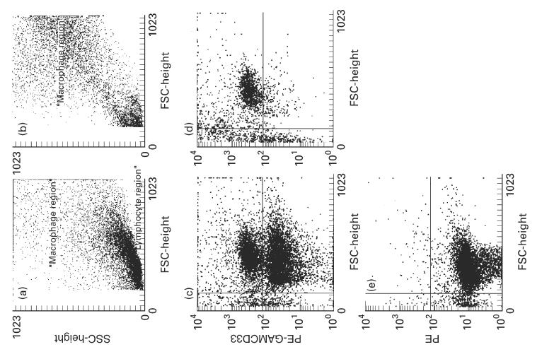
Isolation of CD33+ cells by anti-CD33 magnetic beads. Forward/side scatter characteristics isolated intestinal mononuclear cells before (a) and after (b) two subsequent separation procedures with MACSbeads. The large lymphocyte population has almost completely disappeared in b. (c) Side scatter versus CD33+ cells after one isolation step. CD33+ cells are enriched to about 50% of the cells. (d) After the second separation step > 90% of the intact cells are CD33+. (e) Isotype control showing no non-specific staining.