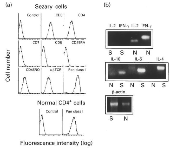
Fig. 1.

Surface phenotype and cytokine mRNA expression pattern of Sézary cells purified from peripheral blood mononuclear cells (PBMC) of the Sézary syndrome (SzS) patient by CD4+ and CD7−selections. (a) The tumour cells and CD4+ cells from PBMC of a normal subject were stained with the indicated FITC-conjugated MoAbs and analysed by flow cytometry. Fluorescein-conjugated mouse IgG MoAb was used as a control antibody. (b) Total RNA was extracted from Sézary cells (S) and PBMC from a normal subject (N). Reverse transcriptase-polymerase chain reaction (RT-PCR) was performed by using mRNA-specific oligodeoxynucleotide primers for IFN-γ, IL-2, IL-4, IL-5 and IL-10 cytokines. Following agarose gel electrophoresis, RT-PCR products were visualized by ethidium bromide.
