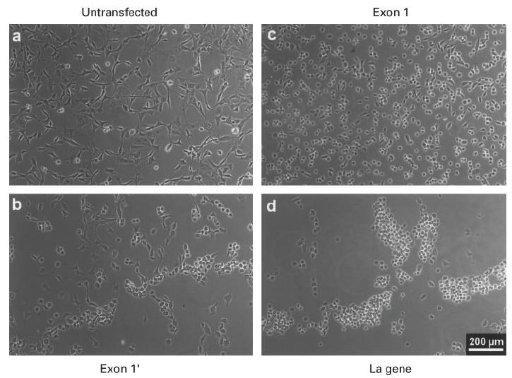
Fig. 5.

Phase contrast analysis of mouse 3T3 cells after herpes simplex type 1 virus infection. The cells were infected with herpes simplex virus type 1 strain ANG. Untransfected mouse 3T3 cells (a), or cells expressing the exon 1′ under cytomegalovirus (CMV) promoter control (b), or cells expressing the human La gene under control of the genuine promoter elements (d), or cells expressing the exon 1 La mRNA under CMV promoter control (c) were fixed 5 h p.i. and phase contrast images were taken.
