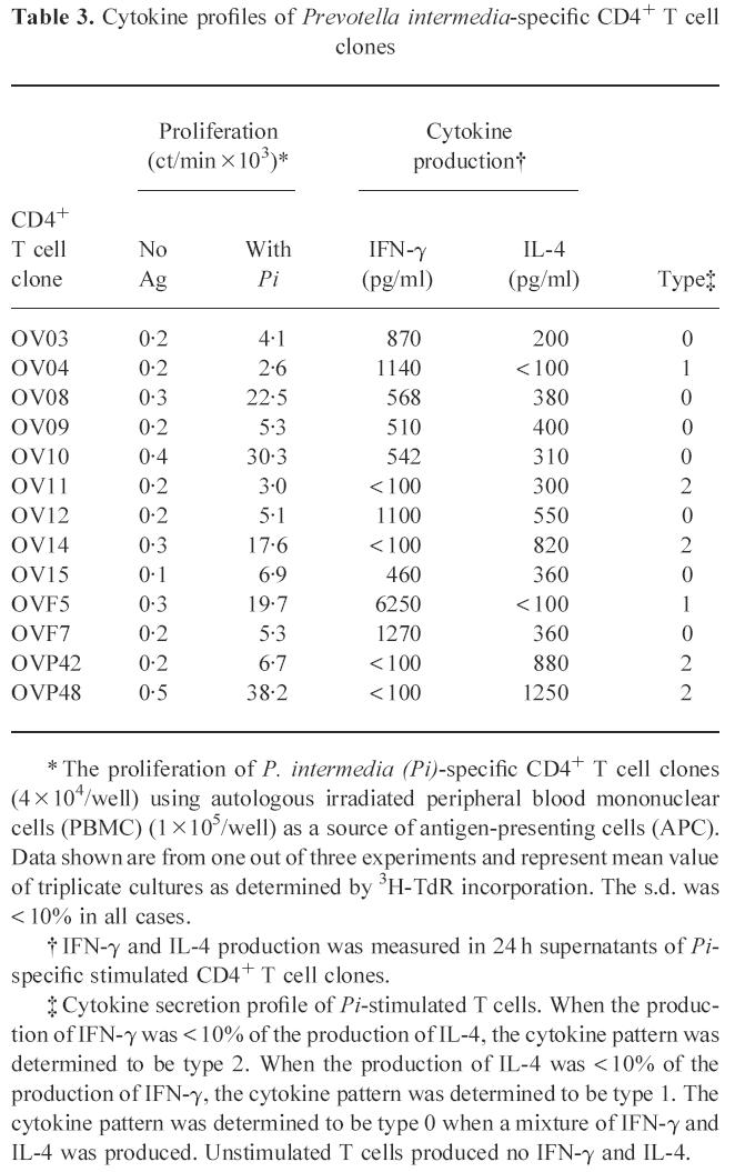
Table 3.
Cytokine profiles of Prevotella intermedia-specific CD4+ T cell clones

* The proliferation of P. intermedia (Pi)-specific CD4+ T cell clones (4 × 10⁴/well) using autologous irradiated peripheral blood mononuclear cells (PBMC) (1 × 10⁵/well) as a source of antigen-presenting cells (APC). Data shown are from one out of three experiments and represent mean value of triplicate cultures as determined by 3H-TdR incorporation. The s.d. was < 10% in all cases.
† IFN-γ and IL-4 production was measured in 24 h supernatants of Pi-specific stimulated CD4+ T cell clones.
‡ Cytokine secretion profile of Pi-stimulated T cells. When the production of IFN-γ was < 10% of the production of IL-4, the cytokine pattern was determined to be type 2. When the production of IL-4 was < 10% of the production of IFN-γ, the cytokine pattern was determined to be type 1. The cytokine pattern was determined to be type 0 when a mixture of IFN-γ and IL-4 was produced. Unstimulated T cells produced no IFN-γ and IL-4.
