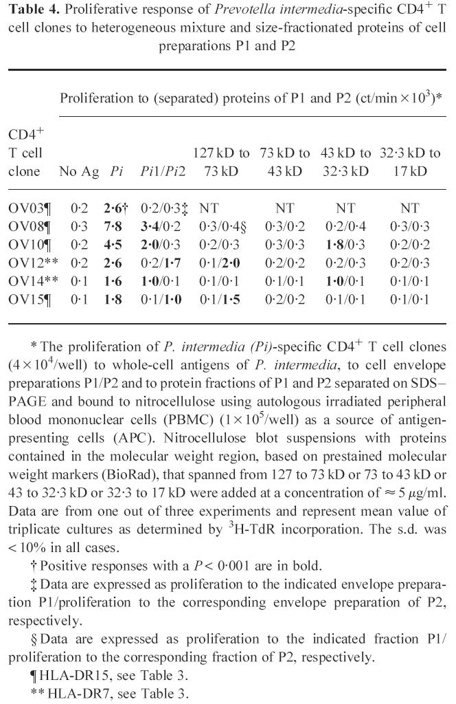
Table 4.
Proliferative response of Prevotella intermedia-specific CD4+ T cell clones to heterogeneous mixture and size-fractionated proteins of cell preparations P1 and P2

* The proliferation of P. intermedia (Pi)-specific CD4+ T cell clones (4 × 10⁴/well) to whole-cell antigens of P. intermedia, to cell envelope preparations P1/P2 and to protein fractions of P1 and P2 separated on SDS–PAGE and bound to nitrocellulose using autologous irradiated peripheral blood mononuclear cells (PBMC) (1 × 10⁵/well) as a source of antigen-presenting cells (APC). Nitrocellulose blot suspensions with proteins contained in the molecular weight region, based on prestained molecular weight markers (BioRad), that spanned from 127 to 73 kD or 73 to 43 kD or 43 to 32.3 kD or 32.3 to 17 kD were added at a concentration of ≈ 5 μg/ml. Data are from one out of three experiments and represent mean value of triplicate cultures as determined by 3H-TdR incorporation. The s.d. was < 10% in all cases.
† Positive responses with a P < 0.001 are in bold.
‡ Data are expressed as proliferation to the indicated envelope preparation P1/proliferation to the corresponding envelope preparation of P2, respectively.
§ Data are expressed as proliferation to the indicated fraction P1/proliferation to the corresponding fraction of P2, respectively.
¶ HLA-DR15, see Table 3.
** HLA-DR7, see Table 3.
