
Fig. 4.
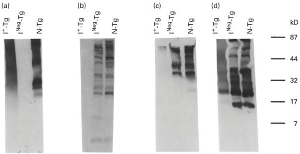
Western immunoblot pattern of immunoreactivity of binding of MoAbs 42C3, 133B1, 41A5 and 137C1 to tryptic peptides of normal Tg (N-Tg), INeg-Tg and I+-Tg. Thirty micrograms of 4-h tryptic peptides of N-Tg, INeg-Tg, and I+-Tg were analysed on a 5–20% SDS–PAGE and the peptides were transferred into a NC membrane. The membrane was then treated with either MoAb 42C3, 133B1, 41A5 or 137C1. (a,b,c,d) The immunoblot patterns of MoAbs 42C3, 133B1, 41A5, and 137C1, respectively. The molecular weights of protein standards are shown on the right.