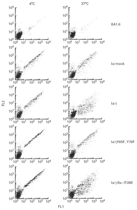
Fig. 2.

Phagocytosis by FcγRIa-expressing IIA1.6 cells. FITC-labelled IgG-opsonized Staphylococcus aureus were incubated with either non-transfected cells, or FcγRIa complex-transfected cells for 45 min at 4°C. Cells were further incubated for 45 min either at 4°C (left lanes) or 37°C (right lanes). Remaining cell surface-bound bacteria were stained with PE-conjugated goat anti-human antibody. FITC (FL1) and PE (FL2) fluorescence of 3500 cells was quantified by flow cytometry, and dot plot diagrams are shown. Experiments were repeated at least four times yielding almost identical results.
