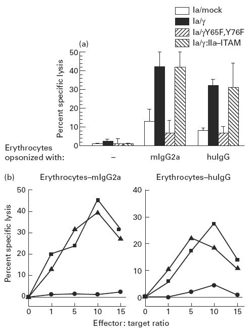
Fig. 3.

(a) ADCC triggered by FcγRIa-transfected IIA1.6 cells. Lysis of erythrocytes was measured by 51Cr release in supernatants after 3 h coincubation of IIA1.6 transfectants with mouse IgG2a (mIgG2a)- or human IgG (huIgG)-opsonized erythrocytes (effector:target ratio of 10). Non-opsonized erythrocytes (−) served as a control. Data represent mean ± s.e.m. of eight individual experiments. ADCC of (mIgG2a- and huIgG)-opsonized erythrocytes by FcγRIa/γ and FcγRIa/γ:IIa–immunoreceptor tyrosine-based activation motif (ITAM)-transfected cells differed significantly from other transfectants and non-transfected IIA1.6 cells (P < 0.05). (b) Erythrocyte lysis at different effector:target ratios by FcγRIa-transfected IIA1.6 cells. IIA1.6 cells expressing either FcγRIa/mock (•), FcγRIa/γ wild type (▪) or FcγRIa/γ:IIa–ITAM (▴) were incubated at different effector:target ratios (indicated on the abscissas) with either mouse IgG2a-opsonized erythrocytes (erythrocytes–mIgG2a) or human IgG-opsonized erythrocytes (erythrocytes–huIgG). Experiments were repeated at least three times, yielding essentially identical results.
