
Fig. 2.
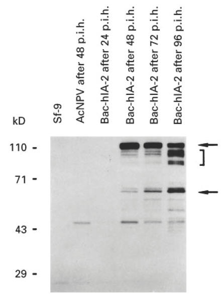
Time course of Bac-hIA-2 expression in Sf-9 cells. At 24, 48, 72 and 96 h post-infection cellular lysates were subjected to 10% SDS–PAGE and immunoblotted with rabbit antiserum to the intracellular domain of human IA-2. The arrows point to the 120-kD full-length human IA-2 protein and the 64-kD IA-2 doublet. The bracket shows degradation products of IA-2. Wild-type baculovirus (AcNPV).