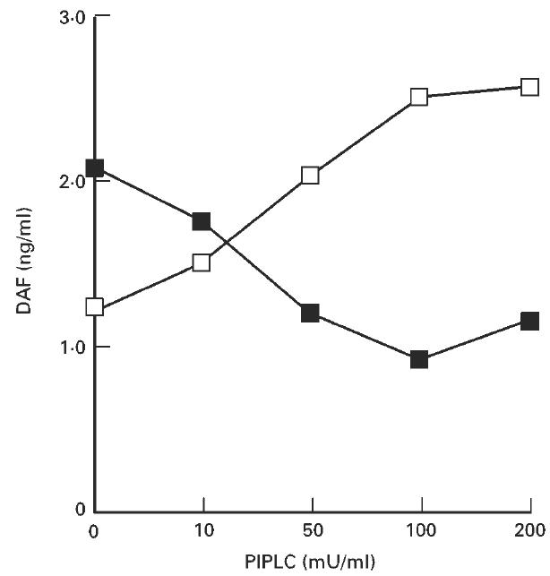
Fig. 2.

Release of DAF from the surface of HT-29 cells by phosphatidylinositol-specific phospholipase C (PIPLC) treatment. HT-29 cells (1.0 × 10⁶ cells) were detached and treated with PIPLC. The amounts of DAF released into the PIPLC solution (□) and in the lysate of remaining cells (×100) (▪) were determined by ELISA. This experiment was repeated twice with similar results.
