Fig. 3.

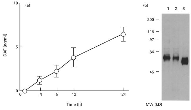
(a) Time-dependent increase in the amount of DAF released into the HT-29 culture supernatant. A supernatant of HT-29 monolayers cultured under standard culture conditions was collected at each time point, and the amount of DAF was determined by ELISA. Values shown are means ± s.e.m. of five experiments. (b) Immunoprecipitation of DAF protein in the culture supernatant of biotin surface-labelled HT-29 cells. The supernatant and cell lysate were immunoprecipitated with Sepharose beads coupled with 1C6 anti-DAF antibody. The Sepharose-bound biotinylated DAF was subjected to electrophoresis and blotted onto a nitrocellulose membrane. The membrane was then treated with horseradish peroxidase (HRP)-labelled streptavidin. Lane 1, membrane-associated DAF; lane 2, DAF spontaneously released into the culture supernatant; lane 3, DAF released with phosphatidylinositol-specific phospholipase C (PIPLC) treatment.
