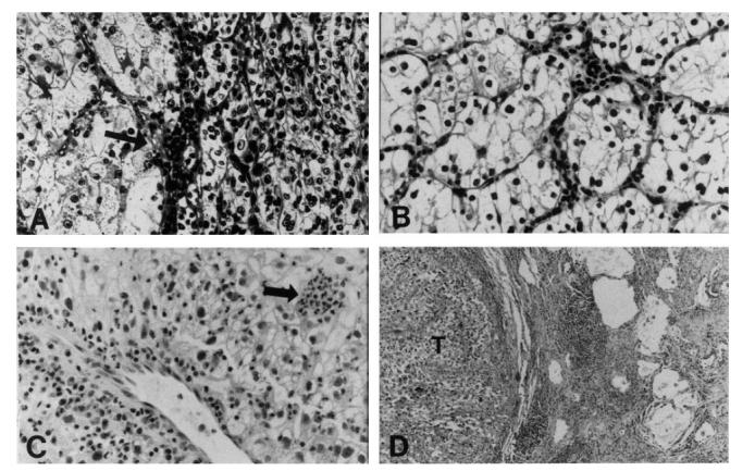
Fig. 2.

Light microscopic sections of the renal tumours and pulmonary nodule. (A) Left kidney tumour showing two growth patterns and scant inflammation in the septum (arrow) (H–E, × 160). (B) Right kidney tumour showing solid nests of clear cells with scant intratumoural inflammation (H–E, × 160). (C) Pulmonary nodule showing perivascular lymphocytes and plasma cells, and clusters of polys amongst the tumour cells (arrow) (H–E, × 160). (D) Zone of lymphoid aggregates and fibrosis at periphery of pulmonary tumour nodule (T) (H–E, × 40).
