
Fig. 3.
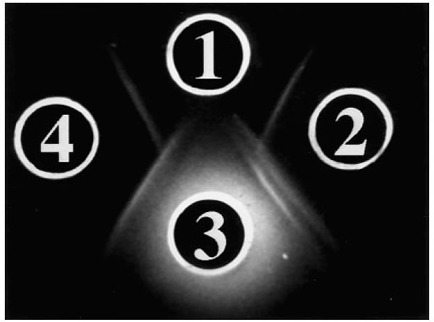
Reactivity of S1 against human liver cytosol fraction and bovine argininosuccinate lyase (ASL), when tested in 0.8% agarose and 3% polyethylene glycol (PEG). Well 1 contains bovine ASL 4 mg/ml, well 3 contains liver cytosol 4 mg/ml, wells 2 and 4 contain S1 diluted 1:24 and 1:16, respectively. The serum in well 2 gives a double line against liver cytosol (well 3) and a single line against ASL (well 1). Of the two precipitin lines formed against liver cytosol, the one nearer well 2 gives a pattern of non-identity with the line formed between wells 2 and 1, while the second line gives a pattern of partial identity. A partial mirror image is seen when well 4, containing S1 at 1:16 dilution, is considered. At this serum dilution the double line between S1 and cytosol is hardly visible.