
Fig. 4.
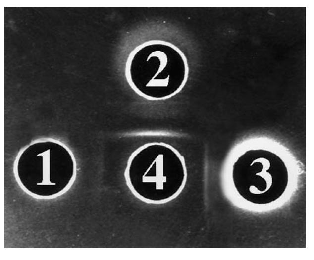
Reactivity of S1 against bovine argininosuccinate lyase (ASL), human ASL, and liver cytosol. Well 1 contains bovine ASL (4 mg/ml), well 2 human ASL (4 mg/ml), well 3 liver cytosol (4 mg/ml). In well 4 there is S1 diluted 1:4. The experiment was performed in 0.8% agarose, 3% polyethylene glycol (PEG). A pattern of identity is given by S1 (4) against human ASL (2) and cytosol (3). The lines between wells 1 and 2 meet at a sharp angle, a pattern compatible with that of partial identity seen in Fig. 3 between the same reactants.