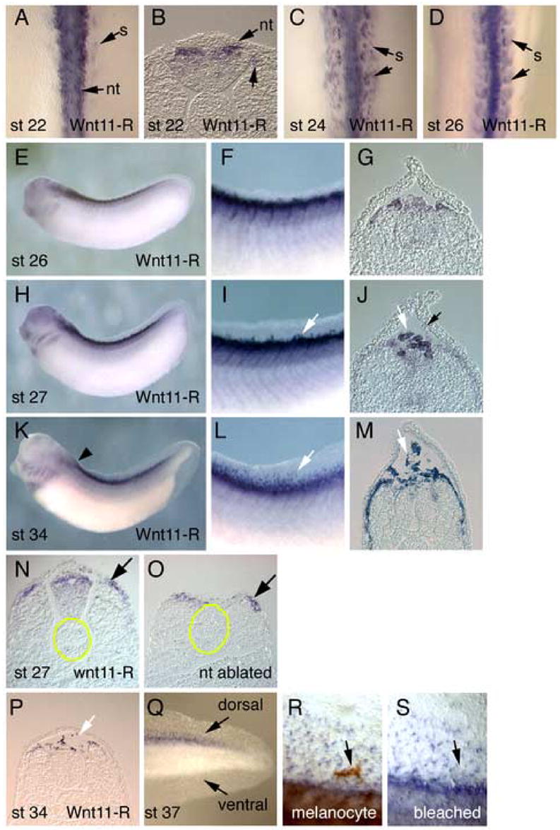
Figure 1.

In situ hybridization analysis of Wnt11-R expression during Xenopus fin development. (A) Dorsal view of St 22 embryo showing Wnt11-R transcripts in the neural tube and in cells at the medial border of the somite. (B) Transverse section through the embryo in (A) showing Wnt11-R expression in the dorsal neural tube. (C) Dorsal view of St 24 embryo showing expression of Wnt11-R in the neural tube and in the somite. (D) Dorsal view of St 26 embryo with Wnt11-R expression in the neural tube and more extensively in the somite. (E) Lateral view of St 26 embryo showing expression of Wnt11-R in the pharyngeal arches and in dorsal tissues of the embryo. (F) Enlarged view of (E) showing expression of Wnt11-R in the dorsal somite. (G) Transverse section showing Wnt11-R transcripts in dorsal neural tube and the dorsal region of the somites. (H) Lateral view of St 27 embryo showing expression of Wnt11-R in dorsal tissues and in cranial NC migrating into the pharyngeal arch region. (I) Enlarged view of (H) showing expression of Wnt11-R in the somite and in individual cells at the base of the dorsal fin (arrow). (J) Transverse section through St 27 embryo showing expression of Wnt11-R in detached cells at the base of the dorsal fin (white arrow). We sometimes observe a small number of cells within the fin that at not expressing Wnt11-R (black arrow). (K) Lateral view of St 34 embryo showing expression of Wnt11-R in the heart, cranial neural crest cells and dorsal tissues. (L) Enlarged view of (K) showing numerous separate stained cells within the fin. (M) Transverse section through St 34 embryo showing Wnt11-R expressing cells dispersed within the dorsal fin core and along the dorsal and lateral surface of the somite. (N, O) Transverse sections through the trunk of a St 25 embryo from which a region of the neural tube has been ablated. For reference, the notochord is outlined in yellow. The section in (N) is located anterior to the region where the neural tube has been removed. Note Wnt11-R expression in the dorsal neural tube and the dorsomedial region of the somite. The section in (O) shows the region where the neural tube is missing. Wnt11-R expression in the dorsal somite (arrow) is equivalent to that in the control section. (P) Transverse section through St 34 embryo at the level indicated by the arrowhead in (K) showing stained cells dorsal to the neural tube. (Q) Lateral view of the tail of St 37 embryo showing Wnt11-R expressing cells within the dorsal fin but not the ventral fin. (R) Magnified view of the dorsal fin of an unbleached St 37 embryo showing Wnt11-R expressing cells, plus a large pigmented melanocyte (arrow). (S) Identical region of the fin shown in (R) after bleaching. Note that the melanocyte does not express detectable levels of Wnt11-R. Abbreviations: nt, neural tube; s, somite.
