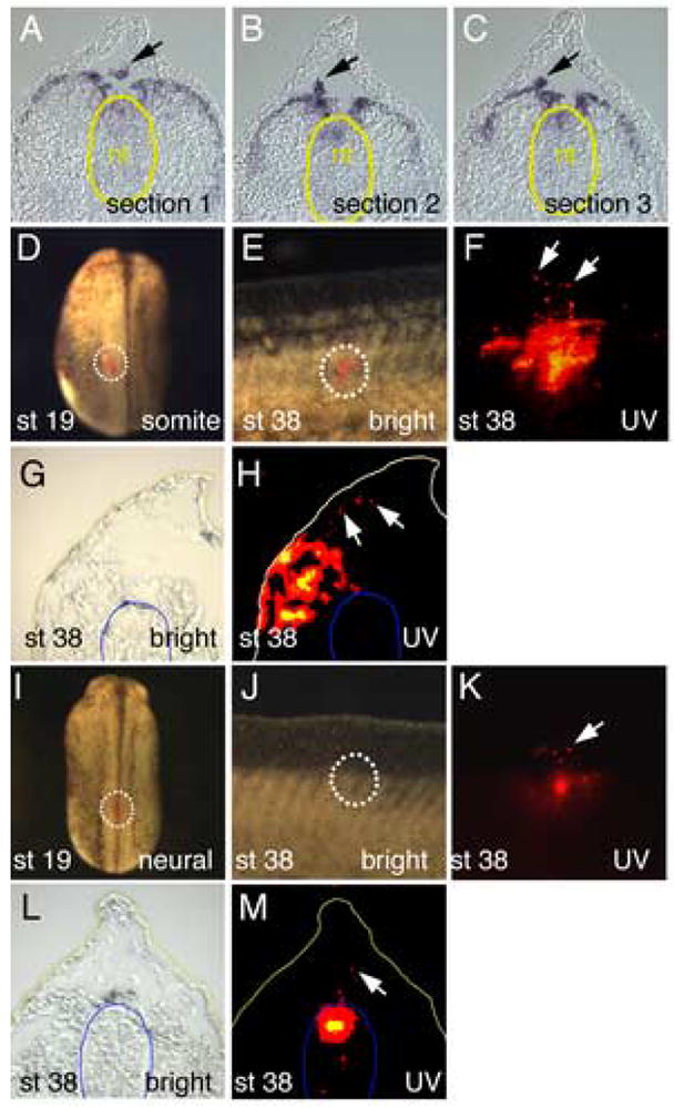
Figure 2.

Lineage tracing of cells migrating into the dorsal fin matrix. (A–C) Serial sections through the trunk region of St 27 embryo stained for Wnt11-R transcripts. Cells moving into the fin (arrows) appear to be detaching from the dorsal somites rather than the neural tube (yellow outline) (D) Embryo at St 19 showing location of DiI label on the dorsal surface of the somite (outline). (E) Merged UV and white light image of the dorsal fin of a DiI labeled embryo at St 38, showing the presence of labeled cells within the fin matrix. (F) UV image of same region in (E) showing presence of DiI labeled cells inside the fin (white arrows). (G, H) Bright field and UV image respectively of frozen section through a DiI labeled embryo at St 38. Cells originating in the labeled somite have migrated into the matrix of the dorsal fin (arrows). The position of the neural tube is outlined in blue for reference. (I). Embryo at St 19 showing location of DiI label on the dorsal surface of the forming neural tube (outline). (J). Merged UV and white light image of the dorsal fin of a DiI labeled embryo at St 38, showing the presence of labeled cells within the fin matrix. (K). UV image of same region in (E) showing presence of DiI labeled cells inside the fin (white arrow). Labeling of the neural tube is faint because it is viewed through the body of the embryo. (L, M). Bright field and UV image respectively of frozen section through a DiI labeled embryo at St 38. Cells originating in the labeled neural tube have migrated into the matrix of the dorsal fin (arrow). The position of the neural tube is outlined in blue.
