Figure 3.

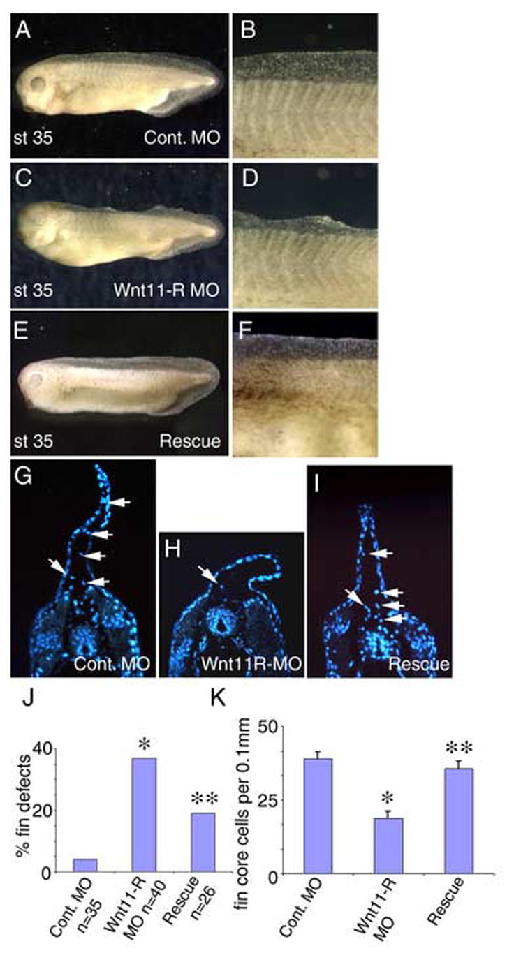
Inhibition of Wnt11-R expression disrupts normal fin development. (A) St 35 embryo injected with 15 ng of control MO showing normal fin morphology. (B) Magnified view of the dorsal fin of the embryo pictured in (A). (C) St 35 embryo injected with Wnt11-R MO showing collapsed appearance of the dorsal fin. (D) Magnified view of the dorsal fin of the embryo pictured in (C). (E) Rescue experiment showing normal dorsal fin development where the embryo was injected with 15 ng of Wnt11-R MO plus 100 pg of Wnt11-R mRNA. (F) Magnified view of the dorsal fin of the embryo pictured in (E). (G–I) UV images of transverse sections through the trunk region of control and MO-treated embryos. Cells within the fin matrix (arrows) can be identified using DAPI nuclear stain. (G) Control. (H) Wnt11-R MO treated embryo showing very few mesenchymal cells within the fin (arrow). Note also the altered morphology of the dorsal fin epidermis. (I) Rescued embryo. The number of cells within the fin is comparable to the control embryo and the dorsal fin morphology is normal. (J) Frequency of embryos showing fin morphological defects in Wnt11-R MO experiments. Statistical difference from control is indicated (*). Coinjection of 100 pg of Wnt11-R mRNA resulted in a reduction in frequency of observed fin defects relative to Wnt11-R MO alone. The rescue was statistically significant (**) using the Chi squared test, p < 0.02. (K) Quantitation of fin core cells per 0.1 mm of dorsal fin in Wnt11-R MO experiments. The decrease in fin core cells in Wnt11-R MO treated embryos was significant, p = 0.00001, by T-Test (Two-Sample Assuming Unequal Variances). The restoration of the number of fin core cells in rescue experiments was also significant (p=0.00009).
