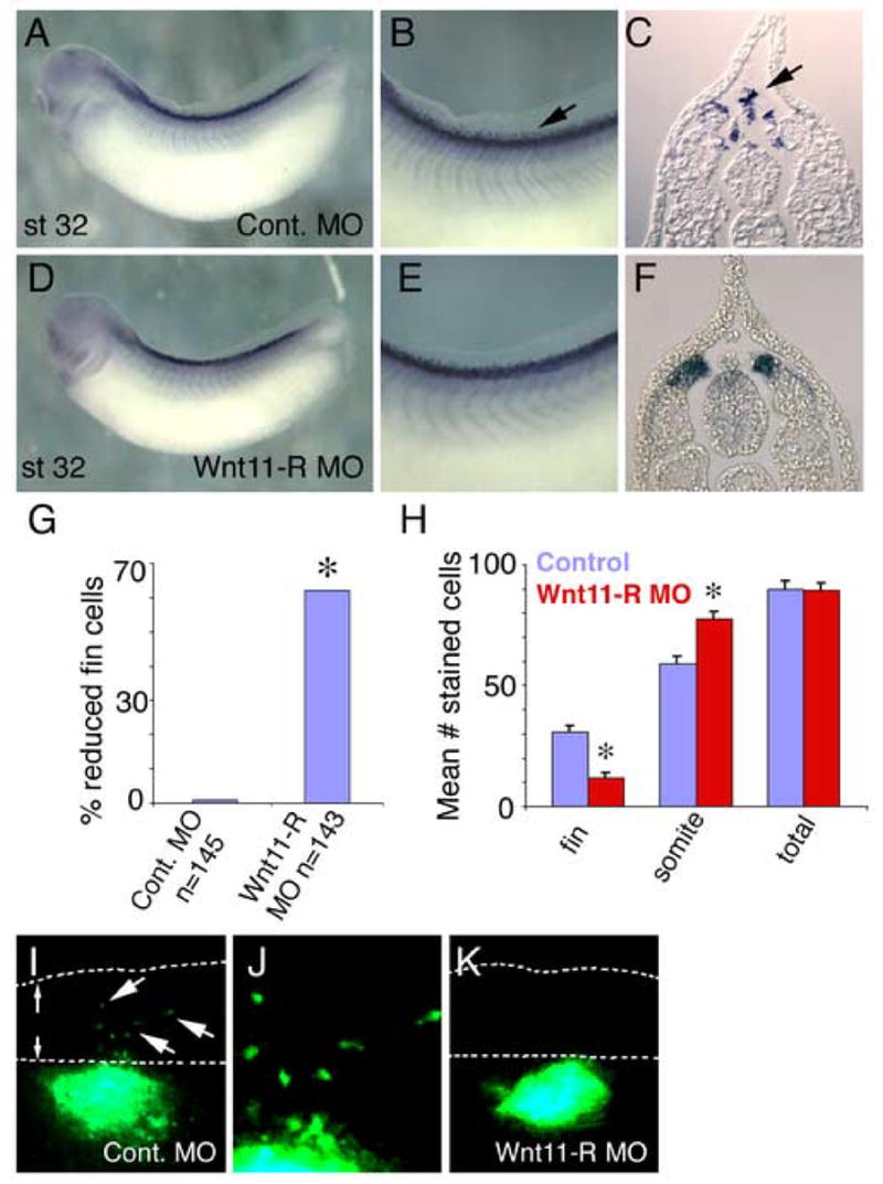
Figure 4.

Inhibition of Wnt11-R expression or non-canonical Wnt signaling results in fin cell migration defects. (A–F) Migratory and premigratory cells were detected by in situ hybridization using Wnt11-R probe. (A) Lateral view of St 32 embryo injected with control MO showing expression of Wnt11-R. (B) Magnified view of the dorsal region of the embryo pictured in (A) showing numerous mesenchymal cells within the dorsal fin (arrow). (C) Transverse section showing Wnt11-R expressing mesenchymal cells detached from the neural tube or somite and entering the dorsal fin. (D) Lateral view of Wnt11-R MO-treated embryo (bilateral injection) showing normal overall morphology and expression of Wnt11-R. (E) Magnified view of the dorsal region of the embryo pictured in (D) showing an absence of separated mesenchymal cells within the fin. (F) Transverse section showing Wnt11-R expressing cells in contact with the neural tube and dorsal somite. (G) Chart showing the percent of embryos with a reduced number of Wnt11-R expressing cells within the fin in MO knockdown experiments. Statistically significant differences from controls are indicated (*). (H) Chart showing quantitation of Wnt11-R expressing cells in 0.1 mm of trunk fin of control and Wnt11-R MO-treated embryos. The number of DAPI stained/Wnt11-R expressing cells within the matrix or attached to the neural tube or the somite is presented, together with total cell number. The decrease in fin core cell number was significant (*) for Wnt11-R MO treatment, (T-Test Two-Sample Assuming Unequal Variances). (I–K) UV images of GFP expressing neural tube implants. (I) GFP labeled cells from control neural tube have migrated from the position of the implant into the dorsal fin. (J) Magnified view of (I) showing isolated mesenchymal cells within the fin. (K) Wnt11-R MO-treated neural tube implant showing absence of detectable mesenchymal cells in the dorsal fin.
