Figure 5.

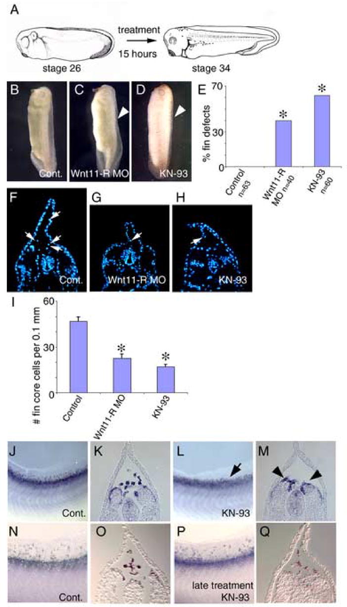
Inhibition of CaMKII activity results in defective migration of cells into the fin. (A) Diagram illustrating the administration of the inhibitor. Embryos were treated with inhibitor from St 26, prior to migration of cells into the fin, until St 34. (B) Control embryo incubated in DMSO carrier medium showing normal dorsal fin development. (C) Wnt11-R MO injected embryo showing disrupted dorsal fin development with a collapsed dorsal fin (arrowhead). (D) CaMKII inhibitor treated (KN-93) embryo showing disrupted dorsal fin development (arrowhead). Note that that dorsal fin is collapsed along the entire length of the embryo. (E) Chart showing the frequency of embryos with fin morphological defects in KN-93 experiments compared to Wnt11-R MO treatments. Statistically significant differences from controls are indicated (*). (F–H) UV images of a transverse sections through the trunk region of control, Wnt11-R MO treated and KN-93 treated embryos. (F) Control embryo showing DAPI-stained fin core cells (arrows). (G) Wnt11-R knockdown embryo showing very few mesenchymal cells within the fin (arrow). (H) KN-93 treated embryo showing very few mesenchymal cells within the fin. Note also the altered morphology of the dorsal fin epidermis compared to control. (I) Quantitation of dorsal fin core cells per 0.1 mm of dorsal fin in Wnt11-R MO and KN-93 experiments. Statistically significant differences from control are indicated (*). (T-Test -Two-Sample Assuming Unequal Variances). (J–Q) Fin mesenchyme cells were detected by in situ hybridization using Wnt11-R probe. (J) Lateral view of the fin region of St 34 control embryo showing numerous mesenchymal cells within the dorsal fin. (K) Transverse section of the embryo in (J) showing Wnt11-R expressing mesenchymal cells detached from the neural tube or somite and entering the dorsal fin. (L) Lateral view of the fin region of KN-93 treated embryo at St 34 showing very few mesenchymal cells within the dorsal fin (arrow). (M) Transverse section of the embryo in (L) showing and absence of fin core cells and Wnt11-R expressing cells attached to the somite (arrowheads). (N) Lateral view of the fin region of St 36 control embryo showing numerous mesenchymal cells within the dorsal fin. (O) Transverse section of the embryo in (N) showing mesenchymal cells within the fin matrix. (P) Lateral view and (Q) transverse section of embryo treated with KN-93 from St 28–36 and assayed at St 36 showing numerous Wnt11-R expressing mesenchymal cells distributed throughout the dorsal fin.
