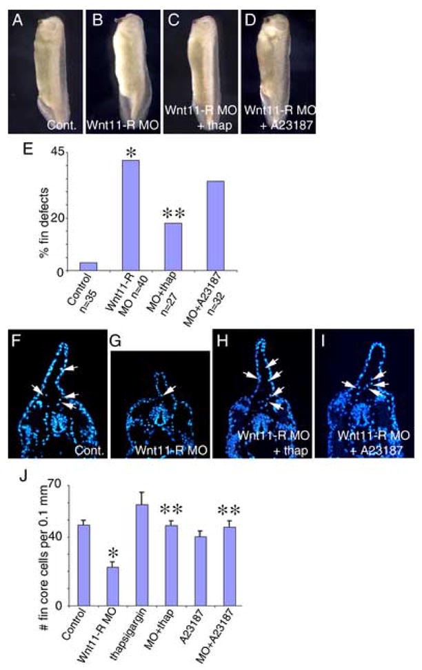
Figure 6.

Activation of calcium signaling rescues dorsal fin development in Wnt11-R knockdown embryos. Calcium signaling was activated using thapsigargin or A23187 for 1 hour at St 26–27 in control MO or Wnt11-R MO-treated embryos. Embryos were subsequently cultured for 20 hours in fresh medium until St 35. (A) Control MO treated embryo showing normal fin development. (B) Wnt11-R MO treated embryo showing defective dorsal fin development. (C) Wnt11-R MO embryo treated with thapsigargin showing normal fin development. (D) Wnt11-R MO embryo treated with A23187 showing normal fin development. (E) Quantitation of dorsal fin morphological defects in Wnt11-R MO embryos (*) and rescue of Wnt11-R MO embryos treated with thapsigargin (**) and A23187. (F–I) UV images of transverse sections through the trunk region of DAPI stained embryos. (F) Control embryo showing numerous DAPI-stained cells within the fin matrix (arrows). (G) Wnt11-R knockdown embryo showing few DAPI-stained cells within the fin matrix. (H) Thapsigargin treated Wnt11-R knockdown embryo showing numerous stained cells within the dorsal fin core. (I) A23187 treated Wnt11-R knockdown embryo showing numerous stained cells within the fin core. (J) Quantitation of fin core cells in 0.1 mm trunk region of experimental embryos. Note the statistically significant decrease in fin cell number in Wnt11-R MO treated embryos (*) and the rescue of fin cell number in Wnt11-R MO embryos treated with thapsigargin or A23187 (**).
