
Fig. 7.
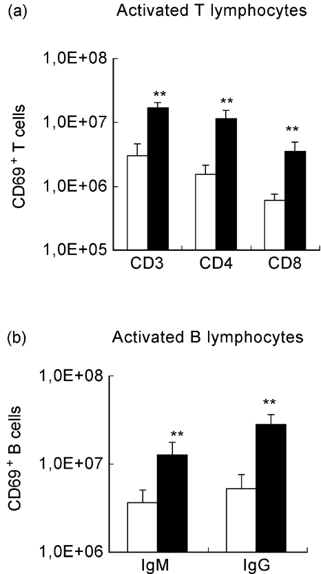
Activation state of lymphocytes from NOD mice after mycobacterial infection. NOD mice were intraperitoneally infected with 108 Mycobacterium avium bacilli or injected with the vehicle of the bacteria. Cell acquisition was performed gating each subset of lymphocytes (CD3+, CD4+, CD8+, IgM+, IgG+) and the percentage of CD69+ cells was determined. The number of CD69+ lymphocytes was derived from the percentage of CD69+ cells and the numbers of splenic CD3+, CD4+, CD8+, IgM+ and IgG+ lymphocytes. Mycobacterial infection induced significant increases in the numbers of activated, CD69-expressing T cells (both CD4+ and CD8+) and activated CD69+ B cells (expressing either surface IgM or IgG). 1,0E + 08 = 108. n = 8. □, Control; ▪, M. avium.