
Fig. 1.
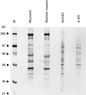
Coomassie blue-stained SDS–PAGE containing human cellular (epithelial cells: A 431; endothelial cells: HUVEC) and tissue-derived (heart, skeletal muscle) protein extracts. M represents the molecular weight marker. Molecular weights are displayed in kD on the left.