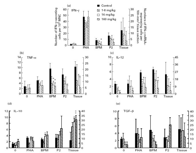
Fig. 6.

Numbers of cells expressing mRNA for IFN-γ (a), tumour necrosis factor-alpha (TNF-α) (b), IL-12 (c), IL-10 (d) and transforming growth factor-beta (TGF-β) (e) per 105 lymph node mononuclear cells (MNC) from experimental autoimmune neuritis (EAN) rats treated with Linomide (1.6, 16 and 160 mg/kg per day) and PBS-treated control rats, after ex vivo culture without antigen or mitogen, and in the presence of phytohaemagglutinin (PHA), bovine peripheral nerve myelin (BPM) or P2 peptide (left ordinate), and per 100-mm2 tissue section of sciatic nerve (right ordinate), obtained on day 27 post-immunization. mRNA expression was detected by in situ hybridization with 35S-labelled synthetic oligonucleotide probes. Bars indicate 1 s.d. P values refer to comparisons between EAN rats treated with Linomide and PBS-treated EAN control rats. *P < 0.05; **P < 0.01.
