
Fig. 1.
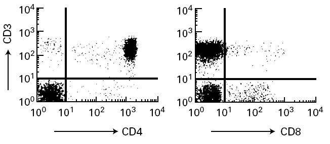
Flow cytometric analysis of patient's peripheral T cells. Patient's peripheral blood mononuclear cells (PBMC) were stained with FITC-labelled anti-CD3 and PE-labelled anti-CD4 or anti-CD8. Stained cells were analysed as described in Patient and Methods.