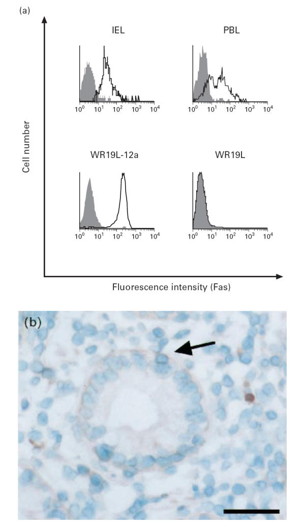
Fig 1.

Expression of Fas protein on freshly isolated colonic intestinal intraepithelial lymphocytes (IEL). (a) IEL and peripheral blood lymphocytes (PBL) were stained with PE-conjugated anti-human CD3 MoAb and FITC-conjugated anti-human Fas MoAb (UB2) (open histograms) or control MoAb (filled histograms), and CD3+-gated cells were analysed by FACSCalibur. Mouse T lymphoma cell line (WR19L) and its human Fas cDNA transfectant (WR19L-12a) were used as negative and positive control, respectively. Data from a representative donor out of five are shown. (b) Normal colonic specimen was stained with UB2. Fas (arrow) is detected in a lymphocyte located adjacent to and slightly deeper than the lining of epithelial cells. The scale bar represents 50 μm.
