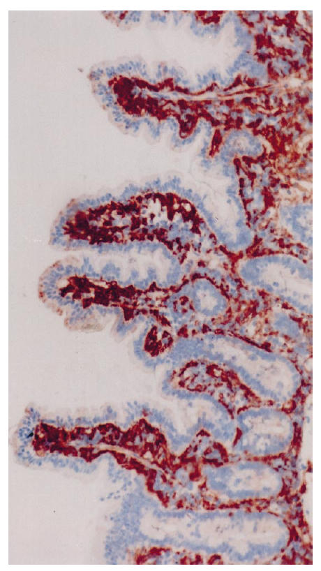
Fig 3.

Biopsy specimen from a control patient treated with antibodies to HLA-DP, original mag. ×10. Only a few positive granules are seen in the apical parts of the epithelial cells at the tip of villi; on average, 1% of the area of epithelial cells was positive.
