
Fig. 2.
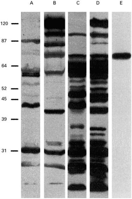
Lanes A, C and B, D, E. SDS–PAGE gel-separated Helicobacter pylori proteins from strains MO19 (Type II) and CCUG 17875 (Type I), respectively. Immunodominant proteins were detected with human sera from an H. pylori-infected individual (lanes A, B), and with bovine colostrum/milk IgG antibodies from cows immunized with strains CCUG17875 and 17874 (lanes C, D) (successfully used in a clinical trial [31]), and with rabbit antisera from immunization experiments with a 20 amino acid synthetic peptide, based on the published 78-kD BabA adhesin sequence (lane E). The top band in lanes B and D corresponds to the 125-kD CagA protein, a member of the cag-PAI (Pathogenicity Island) associated with the more virulent Type I H. pylori strains, and absent from the Type II strains, i.e. strains less associated with acid peptic disease (lanes A, C).