
Fig. 1.
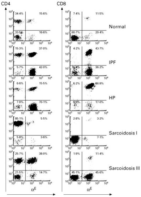
Representative flow cytometric profiles of αE expression on CD4+ and CD8+ bronchoalveolar lavage fluid (BALF) lymphocytes from normal individuals, idiopathic pulmonary fibrosis (IPF), hypersensitivity pneumonitis (HP), sarcoidosis chest radiographic stages I and III. FACS-generated dot plot projections showing fluorescence intensity for gated BALF lymphocytes dual labelled by FITC-conjugated anti-αE (b-ly7) (abscissa, log scale) and PE-conjugated anti-CD4 (Leu-3a) or anti-CD8 (Leu-2a) antibodies (ordinate, log scale).