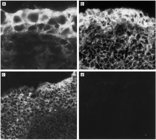
Fig. 2.

Immunostaining for the complement inhibitor membrane cofactor protein (MCP; CD46) in the middle ear mucosa of three different patients with otitis media with effusion (OME) (a–c). The expression of MCP depends on the degree of the epithelial hyperplasia which varied in thickness from two to several cell layers. MCP is expressed throughout the epithelium but the expression is strongest on the two to three outer cell layers. No staining could be detected in the controls stained with the non-specific mouse MoAb AF1 (d). (Mag.: a, × 1150; b, × 630; c–d, × 460.)
