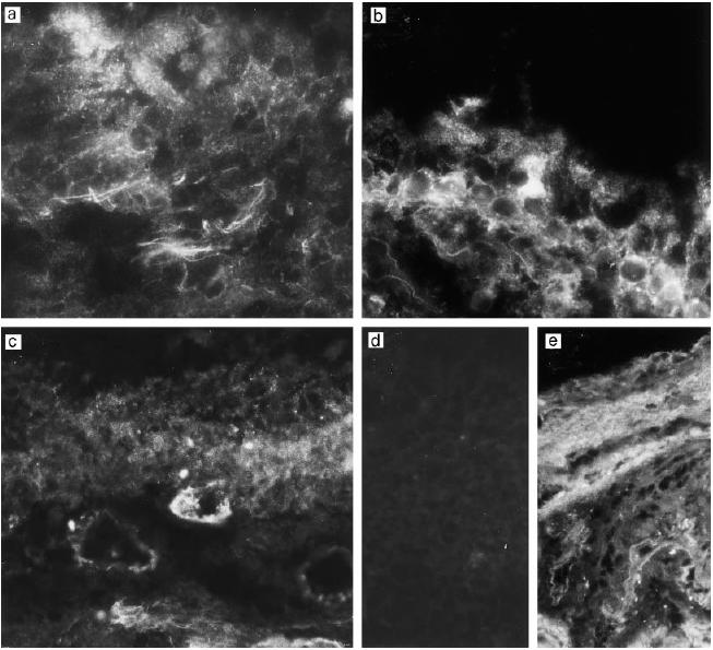
Fig. 3.

Immunostaining of the middle ear epithelium of otitis media with effusion (OME) patients for decay-accelerating factor (DAF, CD55). Samples from three different patients (a–c) show expression of DAF in connective tissue filaments, weakly in the epithelium (a,b) and occasionally in the capillary endothelia (c). No staining is seen in a control stained with the non-specific MoAb AF1 (d). In normal middle ear epithelium the staining pattern for DAF (e) is similar to that in OME patients, except that very little staining is seen in the epithelium. (Mag. × 460.)
