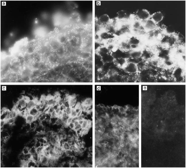
Fig. 4.

Immunostaining of middle ear biopsy specimens for the membrane attack complex inhibitor protectin (CD59). Very strong expression can be seen in samples from three different patients with otitis media with effusion (OME) (a–c). Note the granular pattern of staining (a,b). In OME patients some of the CD59 appears to be peeled off from the outermost cells of the middle ear epithelium (b). The surface of the normal middle ear epithelium also stains positively for CD59 (d). No immunostaining could be detected when the biopsies were stained with a non-specific antibody (e). (Mag.: a,b, × 690; c, × 630; d,e, × 460.)
