
Fig. 2.
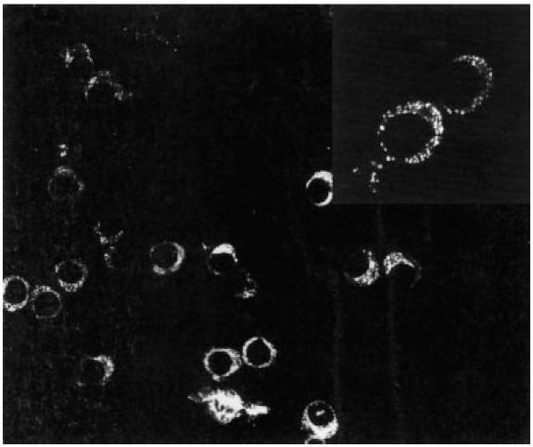
Expression of perforin by tumour-infiltrating lymphocytes (TIL) by indirect immunofluorescence on cytocentrifuged preparations. CD4+ clone PI-H (derived from a glioma lesion) showed bright cytoplasmic staining in approximately half of the cells; at higher magnification (inset) the cytoplasmic stain shows a granular pattern.