
Fig. 3.
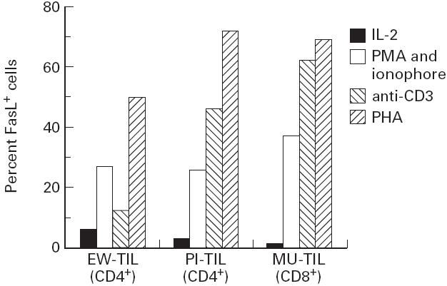
FasL expression by tumour-infiltrating lymphocytes (TIL). Sample 1 is a CD4+ bulk population derived from a melanoma lesion (EW). Sample 2 is a CD4+ bulk population derived from a glioma lesion (PI). Sample 3 is a CD8+ bulk population derived from a melanoma lesion (MU). Cells in IL-2 were stained 6 days after restimulation with phytohaemagglutinin (PHA) and feeder. For other conditions, cells were stained 3 h after restimulation with phorbol myristate acetate (PMA)/ionophore, anti-CD3, or PHA.