
Table 2.
Keratinocytes from lupus patients show enhanced binding of IgG from anti-SS-A/Ro+ sera following UV radiation
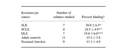
†Each cell strain from each donor was tested at least three times, and the values were summed and expressed as mean ± 1 s.d. of the percentage cells showing positive cell surface IgG binding by flow cytometry analysis. The keratinocytes were irradiated with 100 mJ/cm2 of UVB.*P < 0.001 (versus cells from adult controls, versus cells from neonatal foreskin); P < 0.01 (versus cells from discoid lupus erythematosus (DLE)); **P < 0.01 (versus cells from adult controls, versus cells from neonatal foreskin); ***P < 0.05 (versus cells from neonatal foreskin).