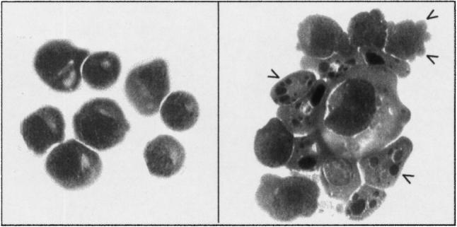
Fig. 1.

Morphological changes that occur during the induction of apoptosis in Jurkat cells after treatment with cyclohexamide. Left panel, untreated cells, right panel apoptotic cells. Arrows indicate blebs, and nuclear condensation and fragmentation. Cytospins were stained with haematoxylin and eosin.
